


Shelties
N° 88 / 03.06.2009 / GB
SHETLAND SHEEPDOG
ORIGIN
Great Britain.
UTILIZATION
Companion dog and Sheepdog.
GENERAL APPEARANCE
Small, long-haired working dog of great beauty, free from cloddiness and coarseness, action lithe and graceful.Outline symmetrical so that no part appears out of proportion to whole. Abundant coat, mane and frill, shapeliness of head and sweetness of expression combine to present the ideal.
IMPORTANT PROPORTIONS
Skull and muzzle of equal length, dividing point inner corner of eye. Slightly longer from point of shoulder to bottom of croup than height at withers.
BEHAVIOUR/TEMPERAMENT
Alert, gentle, intelligent, strong and active. Affectionate and responsive to his owner, reserved towards strangers, never nervous.
HEAD
Head refined and elegant with no exaggerations; when viewed from top or side a long, blunt wedge, tapering from ear to nose. Width and depth of skull in proportion to length of skull and muzzle. Whole to be considered in connection with size of dog.
CRANIAL REGION
Skull : Flat, moderately wide between ears, with no prominence of occipital bone. Topline of skull parallel to topline of muzzle.
Stop : Slight but definite.
FACIAL REGION
The characteristic expression is obtained by the perfect balance and combination of skull and foreface, shape, colour and placement of eyes, correct position and carriage of ears.
Nose : Black.
Lips : Tight with black rims.
Jaws/Teeth : Jaws level, clean, strong with well-developed underjaw. Teeth sound with a perfect, regular and complete scissor bite, i.e. upper teeth closely overlapping lower teeth and set square to the jaws. A full complement of 42 properly placed teeth highly desired.
Cheeks : Flat, merging smoothly into well rounded muzzle.
Eyes : Medium size obliquely set, almond-shape with black rims. Dark brown except in the case of merles, where one or both may be blue or blue flecked.
Ears : Small, moderately wide at base, placed fairly close together on top of skull. In repose, thrown back; when alert brought forward and carried semi-erect with tips falling forward.
NECK
Muscular, well arched, of sufficient length to carry head proudly.
BODY
Back: Level, with graceful sweep over loins.
Croup: Sloping gradually to rear.
Chest: Deep, reaching to point of elbow. Ribs well sprung, tapering at lower half to allow free play of forelegs and shoulders.
TAIL
Set low; tapering bone reaches to at least hock; with abundant hair and slight upward sweep. May be slightly raised when moving but never over level of back. Never kinked.
LIMBS FOREQUARTERS
Forelegs straight when viewed from front, muscular and clean with strong, but not heavy, bone.Shoulders : very well laid back. At withers, separated only by vertebrae, but blades sloping outwards to accommodate desired spring of ribs. Shoulder joint well angled.
Upper arm : Approximately equal in length with shoulder blade.
Elbows : Equidistant from ground and withers.
Metacarpus (Pastern) : Strong and flexible.
HINDQUARTERS
Thigh : Broad and muscular, thigh bones set into pelvis at right angles.
Stifle : Joint has distinct angle.
Hock : Joint clean cut, angular, well let down with strong bone. Hocks straight when viewed from behind.
FEET
Oval, soles well padded, toes arched and close together.
GAIT/MOVEMENT
Lithe, smooth and graceful with drive from hindquarters, covering the maximum amount of ground with the minimum of effort. Pacing, plaiting, rolling, or stiff, stilted, up and down movement highly undesirable.
HAIR
Double; outer coat of long hair, harsh-textured and straight. Undercoat soft, short and close. Mane and frill very abundant, forelegs well feathered. Hindlegs above hocks profusely covered with hair, below hocks fairly smooth. Face smooth. The coat should fit the body and not dominate or detract from the outline of the dog. Smooth-coated specimens highly undesirable.
COLOUR
Sable : clear or shaded, any colour from pale gold to deep mahogany, in its shade, rich in tone. Wolf-sable and grey undesirable.
Tricolour : intense black on body, rich tan markings preferred.
Blue Merle : clear silvery blue, splashed and marbled with black. Rich tan marking preferred but absence not penalised. Heavy black markings, slate or rusty tinge in either top or undercoat highly undesirable; general effect must be blue.
Black and White, and Black and Tan : also recognised colours.
White markings may appear (except on black and tan) in blaze, collar and chest, frill, legs and tip of tail. All or some white markings are preferred (except on black and tan) but absence of these markings not to be penalised. Patches of white on body highly undesirable.
SIZE AND WEIGHT
Ideal height at withers:
Males 37 cm
Females 35,5 cm
More than 2 1/2 cm above or below these heights highly undesirable.
N.B
Male animals should have two apparently normal testicles fully descended into the scrotum.
FAULTS
Any departure from the foregoing points should be considered a fault and the seriousness with which the fault should be regarded should be in exact proportion to its degree and its effect upon the health and welfare of the dog.
Any dog clearly showing physical or behavioural abnormalities shall be disqualified.
16
Junior champions
17
Champions
23
Qualifiers in Agility
10
Health tests
Health
For us health is one of the most important things for breeding, so our Border Collies and Shelties always will be tested about patologies typical of their breed.
Luckily, today we can control some of these diseases genetically, ensuring that our puppies will be free of suffering from that disease controlled by DNA.
The Shetland Sheepdog (Sheltie), we can do for CEA, DM, vWD III, PRA and MDR1.
The hip, elbow dysplasia, Patella and shoulder OCD can not be controlled genetically by the time, we test each dog. In our case, all our dogs are tested and certified by AVEPA and/or OFFA.
Laboratories for genetic testing:
Optigen: (CEA y CL)
www.optigen.com
Laboklin (CEA, CL, TNS, MDR1, vWD III)
www.laboklin.de
University of NSW (TNS y CL)
http://bordercolliehealth.com
Antagene (MDR1, CEA):
www.antagene.com
SVGM (MDR1)
www.vetgenomics.com/es/
Genomia(MDR1, CL, vWDIII)
www.genomia.cz
Offa(DM)
http://www.offa.org/dnatesting/dm.html
Slovgen (CEA, CL, TNS, MDR1, IGS, DM, MH)
http://www.slovgen.sk/
CEA
The CEA (Anomaly of the eye of the Collie) also is well-known like CH because its main manifestation is hipoplasia coroidal (defective development of dark and vascular the layer of the eye located after the retina and whose function is to nourish to this one and to the crystalline).
It is a disease that affects the Border Collie, Rought Collie, Bearded Collie, Shetland and Australian Pastor, and for which is no treatment.
In its more benign manifestation, the dog conserves the vision throughout its life, nevertheless, these units can produce seriously affected descendants. In their more severe manifestation, hemorrhages within the eye can be produced, giving rise serious to a lost one of vision. Usually it is pronounced both to years of age and can affect one or both eyes.
As much the smooth form as the severe one of disease CEA/CH, must to a recesiva mutation in the canine chromosome number 37. There are no indications that nongenetic factors at the time of determining the gravity of the disease take part. The hypothesis that is shuffled, is that there are other certain genes not yet that influence the expression of the gene responsible for the disease.
The disease is easily recognizable in a ophtalmologic examination of the bottom of the eye to 5-8 weeks of life, since the coroides appear pale and fine, is almost transparent, and the blood vessels are appraised easily. Once the retina changes its color of adult (to the rededor of the 3 months of age), the pigment masks the deficiencies in the coroides. The ophtalmologic tests, always before the 3 months of age, allow to determine if the individual suffers or not it disease but we want to make sure that in addition to not suffering it, it will not be transmitted to him to the descendants, it is necessary a genetic test that determines the absence of the mutation.
The only test that allows to determine if a dog is normal, carrier or affected is a genetic analysis that verifies the presence of the mutation in chromosome 37. This test is made in some laboratorios like OPTIGEN, SLOVGEN, LABOKLIN, ANTAGENE…… ; they can certify in writing that certain unit of Border Collie does not suffer the disease and that no of its descendants will suffer it.
Source: Los Trastolillos.
DM (Degenerative Myelopathy)
Degenerative myelopathy is a progressive disease of the spinal cord.
Usually, the disease manifests between 8 and 14 years old. It starts with a loss of coordination (ataxia) in the hind limbs. The affected dog walking staggers, leans over the knuckles or shuffling. At first it may appear at a rear end and then affect the other. As the disease progresses, the limbs become weak and the dog begins to have difficulty keeping up. The weakness gets progressively worse until the dog can not walk. The clinical course can vary from six months to one year before the dog left paraplegic. If symptoms continue a longer period of time, can lead to loss of urinary and fecal continence and eventually also develops on the forelimbs.
A key feature of the DM is that it is not a painful disease.
There are no treatments that have demonstrated a clear efficiency to stop or slow the progression of DM.
The test is provided in laboratories as SLOVGEN, LABOKLIN …
MDR-1 (Multi drug resistance)
The MDR-1 or multidrug resistance gene is responsible for producing a transporter protein (P-glycoprotein) which represents a functional barrier protecting the brain against drugs and other toxins.
When the gene mutates and is not functional, drug substances accumulate in the brain and other organs like the liver and kidneys, they become toxic and cause neurological damage, liver, kidney and even coma and death of the animal.
Some breeds of dogs, among which are the Collie family are more likely to present the mutation. In the United States, approximately three out of four Collies have the MDR-1 gene mutation. The frequency is the same in France and Australia. The mutation of this gene has been found in Shetland, Australian Shepherd, German Shepherd, Bobtail …
Several drugs in common use can pass the barrier for deficiency of MDR-1 gene. The best known are the Ivermectin (antiparasitic) and loperamide (antidiarrheal marketed in Spain and Salvacolina Fortasec).
Using a test can determine whether a specimen has the functional gene, in which case you can administer these medications as normal, or mutated, then you must find an alternative treatment.
Laboratories like SLOVGEN, LABOKLIN, ANTAGENE……provide this test.
Result of DNA test drug sensitivity gene status of the genetic abnormality transmission
Homozygote normal 2 normal copies of the gene MRD1, normal doses of ivermectin, loperamide and other drugs do not cause allergic reactions. They don´t transmit the gen to their offspring.
Heterozygote mutant 1 defective and 1 normal copy of the gene MRD. High doses of ivermectin and normal doses of loperamide and other drugs may be toxic. They can transmit the gen to their offspring. Statistically 50% of their offspring can carry the mutated gen.
Homozygous mutant 2 defective copies of the gene MRD1 High doses of ivermectin and normal doses of loperamide and other drugs are highly toxic. They transmit the gen to their offspring. Statistically 100% of their offspring carry the mutated gen.
Source: Los Trastolillos
vWD‐III Von Willebrand
Von Willebrand disease Type III is a very severe disease, affected animals don´t produce von Willebrand factor protein in their blood. The absence of this protein makes them more susceptible to severe and abnormal bleeding. This can lead to life-threatening situations in cases such as wounds, castration or sterilization. As an autosomal recessive, the Shetland Sheepdogs who are carriers of the disease don´t show clinical signs of vWD, but they can pass the mutated gene to their offspring. If they are not tested, the result is the increase of affected animals.
Although a significant frequency of vWD in Shelties, there is no effective treatment for the disease. Responsible breeders have sought to use protein-based analysis vWD, but has not succeeded in reducing the frequency of the disease. There are too many variables, such as menstrual cycle or thyroid function, which produce variations in results. Therefore, breeders have been unable to fight the disease until now using crosses aimed to reduce the incidence of vWD in future generations.
The test for vWD Type III identifies the presence of the mutation responsible of vwD III and dogs classified as free, CARRIER and AFFECTED.
Frequency of the mutation
Genotype vWD
vWD Type III Free Carrier Affected
Shetland Sheepdog 90.3% 9.4% 0.3%
PRA
Progressive Retinal Atrophy (PRA) is a genetic, degenerative retinal disease that leads to blindness. Sadly enough, it is being discovered in Shetland Sheepdogs and with every PRA-affected Sheltie many carriers surround it.
In the Sheltie and most every other breed studied, PRA is a non-linked, recessive disorder. The recessive PRA gene is a “hidden monster.” It can hibernate for generations. But when paired with a recessive PRA gene from another dog, it comes out of hiding and effects some, if not all of the puppies in that litter. Unfortunately, a Sheltie may be old enough to breed before a veterinary ophthalmologist can detect the disease. And so PRA is passed on to more Shelties. PRA is a GENETIC DISEASE and THERE IS NO CURE.
But for Shetland Sheepdog there is a possibility for research, tell Dr. Simon Petersen-Jones Assistant Professor, Comparative Ophthalmology Department of Small Animal Clinical Sciences Michigan State University. His group developed the first DNA test for PRA. This was in the Irish setter breed. They have also identified the cause of PRA in the Cardigan Welsh corgi and developed a DNA-based test for that form of PRA which has enabled breeders to ensure that no more affected dogs are produced. The test also allows the use of carrier dogs in breeding, this saving the good features from such dogs while ensuring that PRA does not occur.
The Norwegian Shetland Sheepdog club has researched the PRA in this breed and It has provided us the tool to breed more safely taking into account the PRA test . Despite it was already being monitored by us by testing opur Shelties regularly through ECVO certificates, we have the opportunity now to discard their genetic component.
Information based provided in Norwegian Shetland Sheepdog club: http://www.nssk.no/PRA/PRA.html
OCD (Osteochondritis dissecans)
Osteochondritis dissecans (OCD) is considered a hereditary disease, common in large breeds and rapid growth.
The higher incidence of OCD in Border Collies would be expected for its size seems due to their behavior: high energy, athletic ability, stamina and quick reflexes, coupled with the abrupt changes in speed and direction may predispose them to trauma and stress on the joints than most similar sized breeds do not usually experience.
The condition is detected in the pups at the age of 4-9 months but can also be seen in older dogs. Most affected individuals develop clinical signs less than a year old. It seems to be twice as common in males than in females
The shoulder is the most common site of involvement, but can also be seen in the elbow, knee, hock, spine or other joints. Approximately one third of OCD cases, the disease is bilateral (both joints).
It is believed that OCD is caused by a problem in the growth rate of the joint cartilage, in relation to the underlying subchondral bone, which causes it to become thicker. This thickened cartilage are at risk of rupture, especially in areas subject to trauma, stress and movement, as the flow area of the shoulder joint. Repeated trauma causes separation of a fragment of cartilage.
Due to breakage, joint fluid may come into direct contact with sensitive areas of the underlying bone, now exposed, and can cause pain. At this point usually has a limp in the dog. The bits and pieces, over time, they decompose and are absorbed, or may be fed by the joint fluid and grow to a size larger than the original loose cartilage body.
Possible complications arise when the fragments bind to other areas of the joint or trapped in the tendon sheath, causing irritation, obstruction of the circulation and pain.
The diagnosis is confirmed by radiography of affected joints.
The direct factors are considered involved in the development of OCD are rapid growth and trauma in the joint.
Indirect factors affecting rapid growth include nutrition, hormones, genetic predisposition and the size
Hip displasia
 Displasia of hip is a genetic disease (heredable), congenital (present from the birth), degenerative that originates diverse degrees of arthritis, weakness in the later extremities and pain.
In a healthy joint, the head of femur perfectly fits in acetabulo or cavity to acetabular of the hip. In a coxofemoral joint with displasia, there is an alteration in the surfaces you will articulate (deformation of the cavity to acetabular and the head and neck of fémur). The contact lost between the surfaces you will articulate denominates subluxación; if the head of fémur slides outwards in partial or total form of acetábulo of the hip it denominates luxación.
The first passage in the development of the disease affects the cartilage to articulate of the hip, that loses thickness and elasticity, making difficult the absorption of the load during the movement. This loss of thickness of the cartilage can in the open leave completions nervous of the subcondrial bone causing pain. In an attempt to stabilize the union to diminish the pain, the animal produces new bone and the capsule to articulate engrosa, with which the mobility of troops is reduced.
It is not possible to be predicted when a dog with displasia will begin to show clinical signs of lack of mobility (swaying step, rigidity, cojera, difficulty to raise stairs, etc) due to the pain. There are numerous exogenous factors as the caloric contribution, the level of exercise or the meteorological time that can affect to the severity of the clinical signs and their fenotípica expression (radiográficos changes).
Displasia of hip is a disease of multifactorial origin (inheritance and diverse environmental factors as they can be the feeding, the physical exercise disturbed to early age, excessive weight and hormonal alterations). The heredabilidad is very high and of poligénica dominant transmission (intervention of several genes), which makes difficult to find a test genetic that it determines his presence. The only test on which it is counted at the present time is the radiográfico study.
In the crossings between free individuals of displasia there is a high probability of producing puppies with good hips. The probability is increased based on the number of free ancestors of displasia known in previous generations.
The association of specialistic Spanish veterinarians in small animals (AVEPA) values and certifica the degree of displasia of hip by means of radiological study.
Source: Los Trastolillos
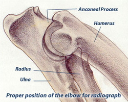
Elbow dysplasia
Elbow dysplasia is a general term used to denote a set of hereditary diseases or injuries (polygenic) that may occur separately or together:
- nonunion anconeus process (UAP).
- fragmented coronoid process of the ulna (FCP)
- osteochondritis dissecans of the medial condyle of the humerus (OCD)
- incongruity of the elbow joint (INC)
<span#ebeff9′” title=”no unión del proceso ancóneo (UAP).”><span#ebeff9′” title=”proceso coronoides fragmentado del cúbito (FCP).”><span#ebeff9′” title=”osteocondritis disecante del cóndilo interno del húmero (OCD).”><span#ebeff9′” title=”incongruencia de la articulación del codo (INC).”> <span#ebeff9′” title=”A los factores genéticos que determinan el grado de severidad hay que añadir otros exógenos como el sobrepeso, el nivel de ejercicio, un aporte excesivo de calcio y vitamina D en la dieta, etc.”>A genetic factors that determine the degree of severity must be added exogenous such as being overweight, the level of exercise, excessive intake of calcium and vitamin D in the diet, etc.<span#ebeff9′” title=”Por eso es tan difícil prever a que edad pueden aparecer los primeros signos clínicos: cojera y caminar rígido del miembro afectado.”>Therefore it is very difficult to predict at what age can receive the first clinical signs of lameness and stiff walk of the affected limb. <span#ebeff9′” title=”Cuando la displasia es bilateral es difícil determinar cual es el miembro más afectado.”>When bilateral dysplasia is difficult to determine which is the most affected member.
<span#ebeff9′” title=”Para realizar un diagnóstico se debe recurrir a las radiografías.”>To make a diagnosis should be made by radiographs.
<span#ebeff9′” title=”El tratamiento suele ser médico (mediante analgésicos y condroprotectores) y, en algunos casos, quirúrgico (eliminando la zona de cartílago afectada).”>Treatment is usually medical (through pain and chondroprotective), and in some cases, surgery (removing the affected cartilage zone).
<span#ebeff9′” title=”En la actualidad no hay ningún test de ADN que permita determinar la presencia de la enfermedad.”>At present there is no DNA test to determine the presence of the disease. <span#ebeff9′” title=”Por eso es tan importante un control radiográfico del mayor número posible de perros.”>Therefore it is very important radiographic control the maximum number of dogs. </span#ebeff9′”></span#ebeff9′”></span#ebeff9′”></span#ebeff9′”></span#ebeff9′”></span#ebeff9′”></span#ebeff9′”></span#ebeff9′”></span#ebeff9′”></span#ebeff9′”></span#ebeff9′”>
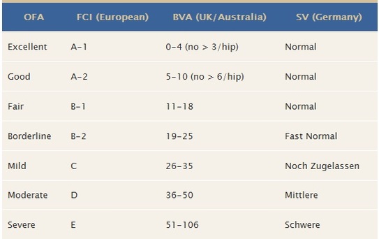
Grading elbows
- Grade 0 Normal: Normal articulation.
- Grade I Elbow Dysplasia: Minimal bone change along anconeal process of ulna (less than 3mm).
- Grade II Elbow Dysplasia: Additional bone proliferation along anconeal process (3-5 mm) and subchondral bone changes (trochlear notch sclerosis).
- Grade III Elbow Dysplasia: Well developed degenerative joint disease with bone proliferation along anconeal process being greater than than 5 mm.
Source: Los Trastolillos
The luxating knee (Patella)
What is Patellar Luxation?The patella, or kneecap, is part of the stifle joint (knee). In patellar luxation, the kneecap luxates, or pops out of place, either in a medial or lateral position.
Bilateral involvement is most common, but unilateral is not uncommon. Animals can be affected by the time they are 8 weeks of age. The most notable finding is a knock-knee (genu valgum) stance. The patella is usually reducible, and laxity of the medial collateral ligament may be evident. The medial retinacular tissues of the stifle joint are often thickened, and the foot can be seen to twist laterally as weight is placed on the limb.
Patellar Luxation CategoriesPatellar luxations fall into several categories:
- Medial luxation; toy, miniature, and large breeds
- Lateral luxation; toy and miniature breeds
- Lateral luxation; large and giant breeds.
- Luxation resulting from trauma; various breeds, of no importance to the certification process.
Numbers 1, 2 and 3 are either known to be heritable or strongly suspected.
Medial Luxation in Toy, Miniature, and Large Breeds
Although the luxation may not be present at birth, the anatomical deformities that cause these luxations are present at that time and are responsible for subsequent recurrent patellar luxation. Patellar luxation should be considered an inherited disease.Clinical Signs
Three classes of patients are identifiable:
- Neonates and older puppies often show clinical signs of abnormal hind-leg carriage and function from the time they start walking; these present grades 3 and 4 generally.
- Young to mature animals with grade 2 to 3 luxations usually have exhibited abnormal or intermittently abnormal gaits all their lives but are presented when the problem symptomatically worsens.
- Older animals with grade 1 and 2 luxations may exhibit sudden signs of lameness because of further breakdown of soft tissues as result of minor trauma or because of worsening of degenerative joint disease pain.
Signs vary dramatically with the degree of luxation. In grades 1 and 2, lameness is evident only when the patella is in the luxated position. The leg is carried with the stifle joint flexed but may be touched to the ground every third or fourth step at fast gaits. Grade 3 and 4 animals exhibit a crouching, bowlegged stance (genu varum) with the feet turned inward and with most of the weight transferred to the front legs.
Permanent luxation renders the quadriceps ineffective in extending the stifle. Extension of the stifle will allow reduction of the luxation in grades 1 and 2. Pain is present in some cases, especially when chondromalacia of the patella and femoral condyle is present. Most animals; however, seem to show little irritation upon palpation.
Lateral Luxation in Toy and Miniature Breeds
Lateral luxation in small breeds is most often seen late in the animal’s life, from 5 to 8 years of age. The heritability is unknown. Skeletal abnormalities are relatively minor in this syndrome, which seems to represent a breakdown in soft tissue in response to, as yet, obscure skeletal derangement. Thus, most lateral luxations are grades 1 and 2, and the bony changes are similar, but opposite, to those described for medial luxation. The dog has more functional disability with lateral luxation than with medial luxation.
Clinical Signs
In mature animals, signs may develop rapidly and may be associated with minor trauma or strenuous activity. A knock-knee or genu valgum stance, sometimes described as seal-like, is characteristic.
Sudden bilateral luxation may render the animal unable to stand and so simulate neurological disease. Physical examination is as described for medial luxation.
Lateral Luxation in Large and Giant Breeds
Also called genu valgum, this condition is usually seen in the large and giant breeds. A genetic pattern has been noted, with Great Danes, St. Bernards, and Irish Wolfhounds being the most commonly affected. Components of hip dysplasia, such as coxa valga (increased angle of inclination of the femoral neck) and increased anteversion of the femoral neck, are related to lateral patellar luxation. These deformities cause internal rotation of the femur with lateral torsion and valgus deformity of the distal femur, which displaces the quadriceps mechanism and patella laterally.
Clinical Signs
Bilateral involvement is most common. Animals appear to be affected by the time they are 5 to 6 months of age. The most notable finding is a knock-knee (genu valgum) stance. The patella is usually reducible, and laxity of the medial collateral ligament may be evident. The medial retinacular tissues of the stifle joint are often thickened, and the foot can often be seen to twist laterally as weight is placed on the limb.
Source: OFA http://www.ofa.org/pl_overview.html
ASMOAN-LASBORRAS
We have Shelties ( Shetland Sheepdog) who will be tested for hip and elbow dysplasia, CEA,MDR1, vWD III, DM and eye diseases
We also take care of the character of our dogs, selecting dogs that make us happy, for us and future owners.
A major task we hope to do with enthusiasm and success.